
- Bioactive Compounds
- By Signaling Pathways
- PI3K/Akt/mTOR
- Epigenetics
- Methylation
- Immunology & Inflammation
- Protein Tyrosine Kinase
- Angiogenesis
- Apoptosis
- Autophagy
- ER stress & UPR
- JAK/STAT
- MAPK
- Cytoskeletal Signaling
- Cell Cycle
- TGF-beta/Smad
- Compound Libraries
- Antibodies
- Bioreagents
- qPCR
- 2x SYBR Green qPCR Master Mix
- 2x SYBR Green qPCR Master Mix(Low ROX)
- 2x SYBR Green qPCR Master Mix(High ROX)
- Protein Assay
- Protein A/G Magnetic Beads for IP
- Anti-Flag magnetic beads
- Anti-Flag Affinity Gel
- Anti-Myc magnetic beads
- Anti-HA magnetic beads
- Poly FLAG Peptide lyophilized powder
- Protease Inhibitor Cocktail
- Protease Inhibitor Cocktail (EDTA-Free, 100X in DMSO)
- Phosphatase Inhibitor Cocktail (2 Tubes, 100X)
- Cell Biology
- Cell Counting Kit-8 (CCK-8)
- Animal Experiment
- Mouse Direct PCR Kit (For Genotyping)
- New Products
- Contact Us
-
 Australia
-
 Austria
-
 Belgium
-
 Brazil
-
 Canada
-
 China
China
-
 Czech Republic
-
 Denmark
-
 Finland
-
 France
-
 Germany
-
 Greece
-
 Hong Kong
-
 Hungary
-
 Iceland
-
 India
India
-
 Ireland
-
 Israel
-
 Italy
Italy
-
 Japan
Japan
-
 Korea
Korea
-
 Luxembourg
-
 Malaysia
-
 Netherlands
-
 New Zealand
-
 Norway
-
 Poland
-
 Qatar
Qatar
-
 Romania
-
 Saudi Arabia
-
 Singapore
-
 Spain
Spain
-
 Sweden
-
 Switzerland
-
 Taiwan
-
 Turkey
-
 United Kingdom
-
 United States
-
 Other Countries
Protein A/G Magnetic Beads for IP
For research use only.
Protein A/G Magnetic Beads for IP use a biological nanosurface technology (S-TEC). Protein A/G is orientated as a coat on the surface of super paramagnetic microspheres with high coating density up to 9.3 × 10¹³ molecules/cm2.

Selleck's has been cited by 247 publications
Advantages
Spinning free, IP (takes <30 minutes) with minimal sample loss.
Background caused by non-specific binding is very low.
Beads contain 9.3×10¹³ molecules/cm2 of protein A/G. Antibody binding capacity up to 0.4-0.5 mg/mL.
Cheap to 73 USD/mL
Price Comparison
Description
Protein A/G Magnetic Beads for IP use a biological nanosurface technology (S-TEC). Protein A/G is orientated as a coat on the surface of super paramagnetic microspheres with high coating density up to 9.3 × 10¹³ molecules/cm2. Compared to other similar immune magnetic beads, Selleck MagBeads™ Protein A/G display more antibody binding sites, therefore during IP, less magnetic beads are used. Non-specific binding is low, enabling Selleck MagBeads Protein A/G to be used in IP conveniently and efficiently. With a large, specific surface area, these beads can greatly shorten the equilibrium antibody and antigen adsorption time, enabling complete antibody antigen adsorption process within 10 minutes, and complete total purification and precipitation in just 30 minutes. This product can be used on a wide variety of samples, such as in cell lysates, supernatants collected from cell secretion, serum, ascites, and other immune antigens.
Specificity
Protein A/G magnetic beads are affinity purification magnetic beads prepared by separately coupling recombinant Protein A and recombinant Protein G to the surface of magnetic beads. Protein A contains four IgG Fc binding regions, while Protein G possesses two IgG Fc binding regions. Protein A/G magnetic beads combine the advantages of both Protein A and Protein G, enabling efficient capture and purification of a wide range of IgG antibodies from various species and subclasses. Compared to using Protein A or Protein G individually, Protein A/G magnetic beads exhibit stronger IgG binding capacity and are less affected by pH fluctuations during the binding process.
Properties
| Concentration | 10 mg/mL |
|---|---|
| Particle size | 100 nm |
| Binding capacity of human IgG | 0.4-0.5 mg/mL |
| Ligand | Recombinant protein A/G |
| Application | Protein Purification, Immunoprecipitation |
| pH stability | 6-8 (Long term) |
Storage (From the date of receipt)
Store at 4°C for 2 years. Don’t freeze in the absence of glycerol.
Protocol
This procedure (Figure 1) offers a general guideline for immunoprecipitation (IP). Optimization may be required for each antibody and target antigen. Protein A/G Magnetic Beads for IP are ideally suited for IP reactions.
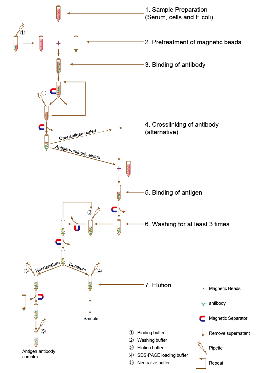
Figure 1 General Protocol for Immunoprecipitation
Recommended buffer examples
| Buffer | Contents (Prepared by customers) |
|---|---|
| Binding buffer | 50 mM Tris, 150 mM NaCl, 0.1%-0.5% detergent (TritonX-100, Tween 20 or NP40), pH 7.5 |
| Wash buffer | 50 mM Tris, 150 mM NaCl, 0.1%-0.5% detergent, pH 7.5 |
| Elution buffer | 0.1 M -0.2 M Glycine, 0.1%-0.5% detergent, pH 2.5-3.1 (or 0.1 M citric acid, 0.1%-0.5% detergent, pH 2.5-3.1) |
| Neutralize buffer | 1M Tris, pH8.0 |
Important Notes before Beginning
1. Before immunoprecipitation, please be sure to carefully read the operating instructions.
2. This product requires use of a magnetic separator.
3. Protein A/G Magnetic Beads should be suspended uniformly before use.
4. Protein A/G Magnetic Beads should be kept in storage solution and prevent dry.
5. Do not freeze or centrifuge MagBeads protein A/G.
6. In order to ensure the best results, please select an antibody with strong specificity.
7. For the IP experiments, different antibodies and antigens will display different binding affinities. Antibody and antigen binding may be altered based on use of binding buffer and washing buffer. Some operator optimization may be necessary.
8. This product is only intended to be used as directed. All other uses are prohibited.
| Species | Antibody class | sProtein A/G | sProtein A |
|---|---|---|---|
| Human | Total IgG | +++++ | +++++ |
| IgG1, IgG2 | +++++ | +++++ | |
| IgG3 | +++++ | + | |
| IgG4 | +++++ | +++++ | |
| IgM | + | + | |
| IgD | - | - | |
| IgA | + | + | |
| IgA1, IgA2 | + | + | |
| IgE | +++ | +++ | |
| Fab | + | + | |
| ScFv | + | + | |
| Mouse | Total IgG | +++++ | +++++ |
| IgM | - | - | |
| IgG1 | +++ | + | |
| IgG2a | +++ | +++ | |
| IgG2b | +++ | +++ | |
| IgG3 | +++ | +++++ | |
| Rat | Total IgG | +++ | + |
| IgG 1 | +++ | +++ | |
| IgG2a | +++++ | +++ | |
| IgG2b | + | +++ | |
| IgG2c | +++++ | +++ | |
| Cow | Total IgG | +++++ | + |
| IgG1 | +++++ | + | |
| IgG2 | +++++ | +++++ | |
| Goat | Total IgG | +++++ | + |
| IgG1 | +++++ | + | |
| IgG2 | +++++ | +++++ | |
| Sheep | Total IgG | +++++ | + |
| IgG1 | +++++ | + | |
| IgG2 | +++++ | +++++ | |
| Horse | Total IgG | +++++ | + |
| IgG(ab), IgG(c) | + | - | |
| IgG(T) | +++++ | + | |
| Rabbit | Total IgG | +++++ | +++++ |
| Guinea pig | Total IgG | +++++ | +++++ |
| Hamster | Total IgG | +++ | +++ |
| Pig | Total IgG | +++++ | +++++ |
| Donkey | Total IgG | +++++ | +++ |
| Cat | Total IgG | +++++ | +++++ |
| Dog | Total IgG | +++++ | +++++ |
| Monkey | Total IgG | +++++ | +++++ |
| Chicken | Total IgG | - | - |
Notes: "+"= weak binding, "+++"=medium binding, "+++++"=strong binding, "-"=no binding
Related Other Products
Tech Support
If you have any other enquiries, please leave a message.
* Indicates a Required Field
Products are for research use only. Not for human use. We do not sell to patients.
©Copyright 2013 Selleck Chemicals. All Rights Reserved.